A normal and typical dorsiventral leaf possesses the following anatomical structure
Upper Epidermis
The internal tissues of the leaf are covered on the upper surface by the upper epidermis. This consists of a single layer of cells with a thick cuticle and usually contains no chloroplasts except in some aquatic plants. Stomata are also usually absent or few except in special cases.
Lower Epidermis
This forms the covering on the lower surface of the leaf and also consists of a single layer of cells with a thin cuticle. It is, however, interspersed by numerous stomata, the guard cells of which contain some chloroplasts.
Mesophyll
The ground tissue lying between the two epidermises is known as the mesophyll. It is differentiated into (a) palisade parenchyma and (b) spongy parenchyma.
- Palisade parenchyma consists of one or two layers of elongated cells with their long axises at right angles to the epidermis. These cells are tightly packed without intercellular space and contain numerous chloroplasts. They are present mainly under the upper epidermis.
- Spongy parenchyma consists of mainly oval, rounded or irregular shaped cells, which contain a few chloroplasts. These cells are very loosely arranged enclosing numerous large interce11ular spaces and air cavities and form the main bulk of the internal tissues of the leaf
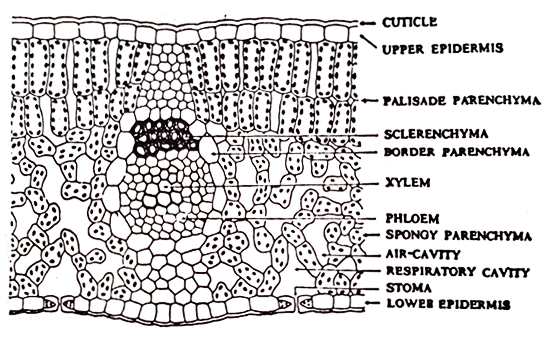
Fig. 23: Internal structure of a dorsiventral leaf. (Adopted from Dutta)
Vascular bundles
They are present in the vascular strands (midrib, veins and veinlets) of the leaf blade, and constitute the conducting tissues of the leaf. Each vascular bundle consists of xylem (always lying towards the upper epidermis) and phloem (always lying towards the upper epidermis) and phloem (always lying towards the lower epidermis) tissues. Xylem consists of various kinds of ~essels (particularly · annular and spiral), tracheids, wood fibres, and wood parenchyma. Phloem consists of sonte narrow sieve tubes, companion cells and phloem parenchyma. Surrounding each vascular bundle there is a bundle sheath, which consists of a compact layer of thin-walled parenchymatous cells. These cells are elongated running parallel with the course of the vascular bundle. Leaves also contain other sclerenchymatous tissues, which do not follow any systematic pattern of distribution. Sometimes the sclerenchyma form patches here and there in the mesophyll, or forms a continuous zone connecting two or more vascular bundles, or occurs as a patch flanking a vascular bundle. Frequently it occurs as a patch flanking a vascular bundle. Frequently is occurs as one or two patches lying associated with the xylem or phloem sometimes it also forms a sheath surrounding a vascular bundle.
