Microscopical measurements or micrometry and drawing to scale are of significant value in the examination of crude drugs, particularly in differentiating an authentic drug from an adulterant, which may have the same tissues, but constituent cells of varying sizes and shapes. Sizes of cells, cellular elements, cell inclusions and other minute structures are measured under the microscope by the use of a micrometer scale inserted into the eyepiece. Tissues, cells and other minute cellular structures are drawn to scale to represent them in their exact natural shape and arrangement by the use of Camera lucida and other similar instruments attached to the microscope.
Measurement of microscopic structures:
Measurements under the microscope are done, as mentioned before, by the use of a micrometer scale called the eyepiece micrometer, fitted inside the eyepiece of the microscope. This micrometer is an arbitrary scale engraved on a glass disc and is divided into 10 equal large divisions, each of which is again divided into 10 equal small divisions. Thus the whole scale has (10 x 10) 100 small equal divisions (see Fig. 73 A). The actual size of each small division on this scale is dependent on the objective magnification · and tube length at which it is being used. In order to determine the size of one small division of the eyepiece micrometer scale. it ts calibrated by the use of a stage micrometer scale. The stage micrometer is a scale of definite length. usually 1 mm long, engraved in a central circle of a glass slide. This scale is also divided into 10 equal large divisions, each measuring 0.1 mm or 100 µm. Each of this large division is again divided into 10 equal small divisions. each measuring 0.01 mm or 10 µm. Thus the stage micrometer scale has also 100 small divisions of 10 µm size (see Fig. 73 B). The marks of the ten large divisions on both the scales are numbered 0 to 100.
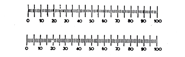
Fig. 73: Micrometer Scales: A, Eyepiece micrometer: B. Stage micrometer
Calibration:
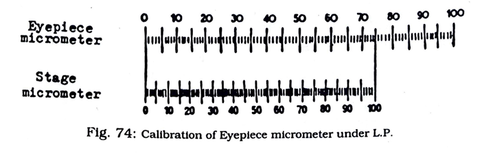
To calibrate the eyepiece micrometer. the required objective (under L.P. or H.P.) is put in position. The Stage micrometer is then focussed in the usual way and the Eyepiece micrometer is superimposed on it in such a way that the O mark on this micrometer coincides exactly with the 0 marks of the Stage micrometer. If calibrating for measurements under low power (L.P.) magnification, a reading is taken at that position on the. Eyepiece micrometer scale to find out the mark which most nearly coincides with the 100 marks of the Stage micrometer scale. For example (see Figure 77), the 75th small eyepiece micrometer division mark coincides with the 100th small stage micrometer division mark. Since one small stage micrometer division is equal to 10 µm, 75 small eyepiece micrometer divisions measure 100 x 10 µm = I 000 µm. Therefore, under this magnification, one small eyepiece micrometer division is equal to 1000 µm + 75 = 13.3 µm.
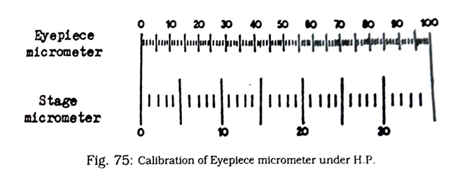
If the Eyepiece micrometer scale is calibrated for measurements under high power (H.P.) magnification, then after focussing the Stage micrometer scale under the H.P. objective a reading is taken at that position on the Stage micrometer scale (now highly magnified) to find out the mark which most nearly coincides with the 100 mark of the Eyepiece micrometer scale (which has not been magnified simultaneously). For example (see Figure 78), the 35th small stage micrometer division mark coincides with the 100th small eyepiece micrometer division mark. Since one small stage micrometer division is equal to 10 µm, 100 small eyepiece micrometer division measure 35 x 10 µm = 350 µm. Therefore, under this magnification, ·one small eyepiece micrometer division is dual to 350 µm + 100 = 3.5 µm.
Thus it is apparent that the size of a small eyepiece m.icrometer division is dependent on the magnification of the objective used, Hence, every time the magnification is changed the Eyepiece micrometer scale has to be freshly calibrated. Once the Eyepiece micrometer is calibrated the Stage micrometer is removed and microscopic measurements are carried out with the Eyepiece micrometer scale, which is still inside the eyepiece, by superimposing it on the object to be measured and counting the number of small divisions covered by the object. The total value of these divisions is the size of the object.
Drawing to scale:
Tissues and other microscopic structures can be drawn to scale and in their exact natural shape and arrangement while being examined under the microscope. For this purpose various types of apparatus are now available commercially. The most commonly used ones of these include the. The Swift-lves camera lucida and the abbe drawing apparatus, The Swift-lves camera lucida (Fig. 79a) is made up of two prisms placed suitable in a dark-coloured metallic casing, and it fits well on top of the eyepiece of the microscope. When in use, light from the object under examination passes direct to the eyes of the
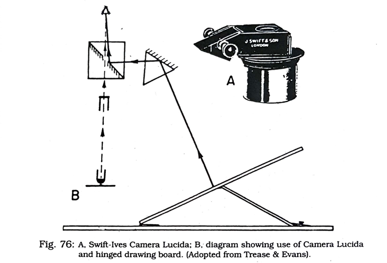
observer through an opening in the silvered surface of the left-hand prism sitting directly on the eyepiece. At the same time light from the suitably positioned drawing paper on the Abbe drawing board (Fig. 79b) and the pencil are reflected by the right hand or side prism and the silvered surface of the left hand prism to reach the observer’s eye simultaneously. Thus the pencil appears superimposed on the object, which may then be traced on the drawing paper.
