External characters refer to the various modifications, artifacts, ramifications and outgrowths that take place on the outer surface of various plant organs. The outer surface of all young plant organs, as mentioned before, is made up of a continuous single layer of closely fitted parenchymatous cells called the epidermis. The epidermis assumes different structures and performs different functions depending on its age and place of occurrence on the plant body. It is also named differently as it matures and occurs on different organs.
For example, when it occurs on leaves and young stem it is called the epidermis; the epidermis of the fruit forms the outermost layer of the epicarp; that of the seed forms the outer layer of the testa; the epidermis of the bark on maturity becomes dead and is replaced by the cork, and that of the young root forms the piliferous layer. Some of the external characters of the plant parts are also produced by the differentiation of the epidermal cells or by the modification of their structures and contents.
The epidermis of the leaves is continuous with that of the stem and is derived from the dermatogen of the apical meristem. Usually, it consists of a single layer of compact cells with occasional stomatal openings, but become multi-layered ion some special cases like the leaves of Piper betle or Ficus elastica.
However, the shape and form of the epidermal cells the nature and structure of their walls; the presence or absence and type of cuticle; the presence or absence of stomata and their type and distribution; the presence of characteristic cell inclusions, and the presence or absence of papillae and trichomes and their forms, types, structures and distribution constitute the external or epidermal characters of the leaves.
The Epidermal Cells
The epidermal cells are tabular and show a complete absence of intercellular spaces except the stomatal pores. They show a great variety in form, giving characteristic pattern in surface view and appearing flattened and square or rectangular in surface view and appearing flattened and square or rectangular in shape in transverse sections. The outer walls are often convex and thickened.
The anticlinal and periclinal walls of the epidermal cells and thickenings on them vary from plant to plant. For example, straight-walled cells are present. in Senna and Coca leaves; wavy-walled cells in Stramonium, Belladonna, and Hyoscyamus; beaded walls In Digitalis lutea and Lobelia inflata.
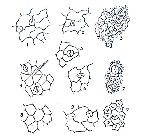
Fig. 24: Epidermal cells and stomata. 1, lower epidermis of Digitalis purpurea with anomocytic stomata; 2 & 3, lower epidermis of Hyoscyamus niger and upper epidermis of Atropa belladonna respectively with anisocytic stomata; 4. Lower epid.ermis of Cassia angustifolia with paracytic stomata; 5 & 6, lower epidermis of Rosmarius of.ficinalis and lower epidermis of Mentha piperita respectively with diacytic stomata; 7, lower epidermis of Pilocarpus jaborandi with actinocytic stomata; 8, upper epidermis of Lobelia inflata; 9, lower epidermis of Digitalis Lan.ala; 10, lower epidermis of Erythroxylon coca. (Reproduced from Trease & Evans).
The Cuticle
The outer walls of epidermal cells are often covered with a waterproof transparent layer of cutting, which is called the cuticle. The cutide is a protective layer and is usually very thin, but with excess deposition of pro-cutin in may produce ridges and become somewhat striated. Cuticle occurs mainly in plants, which conserve water or produce volatile oil. Thick cuticle occurs in Aloe leaf and striated cuticles in Belladonna, Digitalis, and Jaborandi.
Stomata
Stomata arc important epidermal structures of great diagnostic value in the examination of leaf drugs. They are formed by the structural modification of epidermal cells. A stoma consists of two similar cells, called the guard cells, placed with their long axes parallel to each other and having a small intercellular space called the stomatal pore. Through this pore gaseous exchange takes place between the atmosphere and the interior of the leaf. The cells immediately around the stomata are called the subsidiary cells, which may or may not be similar to the other epidermal cells. The type, occurrence and distribution of the stomata in the leaves of different plants vary from plant to plant. They are often characteristic of various leaves and offer important diagnostic characters for the identification of many leaf drugs.
Usually stomata occur both on the upper and lower surfaces of leaves, but their distribution on the two surfaces vary greatly. They may be entirely confined to the lower epidermis as in Ficus, Buchu, Coca and Jaborandi leaves. The floating leaves of aquatic j plants have stomata on their upper epidermis only. Sometimes they are evenly distributed on both surfaces but most commonly ‘ they are more numerous on the lower surface. The number of stomata per square mm of epidermis (stomatal number) and the ratio of stomatal number between the upper and lower epidermis sometimes offer very useful parameters in differentiating between closely related species.
The type of stomata also varies from plant to plant. Based on the form and arrangement of the subsidiary cells, the following five types of stomata may be recognized:
- Anomocytic or ranunculaceous (irregular-celled) type, where the stoma is surrounded by a varying number of subsidiary cells which resemble the other epidermal cells. This type of stomata is found in Digitalis purpurea.
- Anisocytic or cruciferous (unequal-celled) type, where the stoma remains surrounded by 3 or 4 subsidiary cells, of which one is markedly smaller than the others. Anisocytic stomata are found in Stramonium, Belladonna and Hyoscyamus leaves.
- Paracytic or rubiaceous (parallel-celled) type, in which case the stoma has only two subsidiary cells, the long. axises of which are parallel to that of the stoma. Senna leaves possess paracytic stomata.
- Diacytic or caryophyllaceous (cross-celled) type, where the stoma is accompanied by two subsidiary cells, the long axises of which are at right angles to that of the stoma. This type is found in many plants of Labiatae family, e.g. Peppermint, Spearmint and Thyme.
- Actinocytic (radiate-celled) type, where the stoma is surrounded by a number or radiating subsidiary cells arranged along the radii of a circle. This type of stomata is not very common in drug plants.
Cell Inclusions
Cystoliths of calcium carbonate occurs in the epidermal cells of the members of Cannabmaccae spherocrystals of diosmin in Buchu epidermis of senna and Buchu leaves.
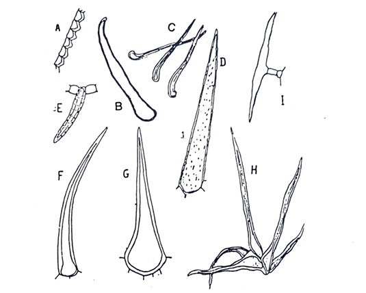
Fig. 25: Unicellular Covering trichomes. A, Papillae of Coca leaf; B-G, Unicellular trichomes: B, Symphytum officinale; C, Thea sinensis; D, Lobelia inflata; E. Senna; F, Ailanthus; G, Comphrey; H, bunch of unicellular trichomes in Hamamelis; I, T-shaped trichome of Artemisia. (After Trease & Evans).
Papillae
Epidermal cells of some plants sometimes extend outwards as dome-shaped or conical projections which are termed as papillae, the epidermis being called papillose. Papillae occur most frequently on the under surface of the leaves of Coca and Cassia obovata.
Trichomes
Trichomes are elongated tubular outgrowths on the epidermal cells of. most leaves, many herbaceous stem, flower, fruit, and seed. They are also called epidermal hairs. The occurrence and forms or the trichomes are very valuable characters for the identification of leaf drugs and detection of adulterants in them. They may be absent from many important Jcaf drugs like Coca, Hemlock and Convallaria, or they may be of rare occurrence as in Buchu and Henna, but many leaves possess them ii~ more or less abundance. Three types of trichomes or hairs are generally recognized;
- Covering or clothing trichomes;
- Glandular trichomes, and
- Hydathodes
And other special types. Anyone type or more than one type of trichomes may occur on the same plant.
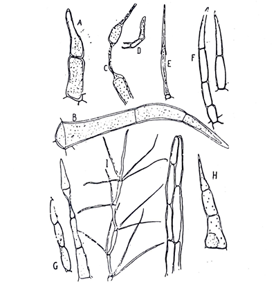
Fig. 26: Multicellular Covering trichomes. A-H, Uniseriate trichomes: A, Datura metel B, Datura stramonium; C, Mentha piperita; D, Thymus vulgaris; E, Plantago lanceolata; F, Hyoscyamus niger; G, Digitalis purpurea; H, Xanthium strumarium· I Multicellular branched trichome of Verbascum thapsus; J, Biscriate trich~mc of Calendula officinalis. (Reproduced from Trease & Evans).
Covering or clothing trichomes
They may be unicellular or multi-cellular. The unicellular trichomes vary greatly. For example, short, conical trichomes occur in Tea and Buchu levels; short, conical and warty trichomes in Senna; large, conical and longitudinally striated trichomes in Lobelia; long, thick-walled trichomes in Ailanthus, and T-shaped trichomes in Artemisia absinthium. They may also occur in a group as in Hamamelis leaf. The multicellular may be uni-, bi- or multiseriate or even branched. The number of cells in the multicellular trichomes may vary from two to many cells. Uniseriate, 2 to 3-celled long covering trichomes are found in Datura and Hyoscyamus; 3 to 4-celled long in Digitalis, and 4 to 5-celled long in Belladonna. Biseriate multicellular trichomes occur on leaves of Calendula officinalis. Multiseriate trichomes are found in Euphorbia hirta. Branched multicellular trichomes may be stellate as in Hamamelis leaves or may be slender and elongated as in Verbascum thapsus.
Covering trichomes function as protective agents, light screen for reducing rate of transpiration, climbing means, water-storing agents and also as seed dispersal agents.
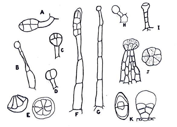
Fig. 27: Glandular trichomes: A & B, Atropa belladonna· C Datura stramonium; D, Digitalis purpurea E Iabiatae plants· Hyoscyamus niger, G & H Primula vulgaris; I, Digitails, lutea; J, Cannabis sativa; K, Artemisia maritima. (Reproduced from Trease & evans).
Glandular trichomes
They usually have uni-or multi-cellular stalks and uni-or multicellular heads. The stalk may be um-or multiseriate and short or long. The glandular trichomes may also be sessile being embedded in the epidermal cells. The type of the glandular trichomes may be characteristic of a plant family or genus, e.g., glandular trichomes with short unicellular
stalks and multicellular rounded or oval heads are found in the Solanaceae and sessile glandular trichomes with multicellular · heads, in which the individual cells radiate, are characteristic of the family Labiatae. However, most glandular trichomes arc multicellular, the simplest type having a uniseriate stalk with a single spherical glandular cell at the apex. This type of trichomes may be found in Digitalis and A Belladonna leaves. But most glandular trichomes of Digitalis purpura have a unicellular stalk and unicellular head. The glandular trichomes of the plants of the Compositae family have short biseriate stalks and bicellular glands. Trichomes having a multiseriate cylindrical stalk and a capitate rosette of secreting cells are found in Cannabis.
Functions of the glandular trichomes include secretion and accumulation of poisonous substances, volatile oils, resins, mucilage, etc
Hydathodes
Hydathodes are special structures formed by the modification of some epidermal cells. Some of them resemble the stomata in structure but do not contain chloroplasts and others resemble the glandular trichomes which are found on the leaves of Piper betle. Their main function is absorption or secretion of water.
There are a large number of leaves, which constitute pharmaceutically useful drugs and commercially important commodities. Some of the important. leaf drugs arc described below in the form of Monographs as reprcscnlat.ivc examples for the students so that they can also describe other leaf drugs in the same way.